M phase is relatively brief and consists of nuclear division (mitosis) and cytoplasmic division (cytokinesis). In mitosis, replicated chromosomes divide, leading to identical daughter nuclei with the same number of chromosomes and the same genetic composition as the parents. In human cells, which have 23 pairs of chromosomes, the number of chromosomes (2n = 46) remains unchanged from the beginning till the end of mitosis.
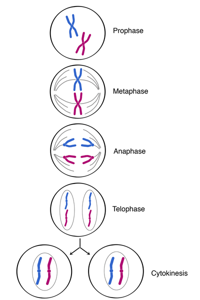
The phases are in the following sequence: prophase, metaphase, anaphase, and telophase (see figure below). The movement of chromosomes is facilitated by a structure called the mitotic spindle, which consists of microtubules and associated proteins. Spindles extend from centrioles on each of the two sides (or poles) of the cell, attach to the chromosomes and align them, and pull the sister chromatids apart.
Chromosomes are usually visible under light microscope. We can determine the number of chromosomes [and chromatid(s) per chromosome] for each stage in mitosis.
Prophase
Chromatin in the nucleus begins to condense and becomes visible under the light microscope as chromosomes, each with two chromatids that are held together at the centromere. The nuclear envelope breaks down. Centrioles begin moving to opposite ends of the cell, and microtubules extend from the centrioles and begin to attach to the centromeres of chromosomes. Eventually, the microtubules extending from centrioles on opposite poles of the cell attach to every centromere and develop into spindle fibers.
Metaphase
By growing on one end and shrinking on the other, spindle fibers align the chromosomes along the middle of the cell nucleus, approximately equidistant from the spindle poles. This imaginary line where the chromosomes are aligned is called the metaphase plate and is equidistant from the centrioles, which are on opposite poles of the cell.
Anaphase
After all of the chromosomes are aligned on the metaphase plate, each pair of sister chromatids splits at the centromere, separates and moves along the shortening spindle fibers to opposite sides of the cell. Now, the number of centromeres and chromosomes within the cell is doubled. Note that each separated chromosome has only one chromatid.
Telophase
Chromosomes, each with one chromatid, arrive at opposite poles of the cell, and a new nuclear membrane forms around each of the two new daughter nuclei, which are identical to each other. The spindle fibers begin to disperse and the chromosomes decondense; chromosomes are no longer visible under the light microscope. Notice that the number of centromeres and chromosomes within each of the two new cells is identical to the parent cell before it underwent mitosis.
Cytokinesis
Although the nucleus divides during mitosis, the cytoplasm does not. Following mitosis, cytokinesis is the process where the cytoplasm of a eukaryotic cell is divided to form two identical daughter cells. In animal cells, the center of the cell contracts, pinching the cell into two daughter cells, each of which contains a nucleus with a complete genome.