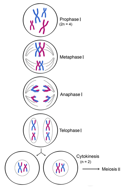
The steps leading up to meiosis are similar to those of mitosis – the centrioles and chromosomes are replicated. The amount of DNA in the cell has doubled, and the ploidy of the cell remains the same as before, at 2n. In meiosis I, the phases are analogous to mitosis: prophase I, metaphase I, anaphase I, and telophase I (below figure). Meiosis I proceeds directly to meiosis II without going through interphase.
Meiosis I is unique in that genetic diversity is generated through crossing over and random positioning of homologous chromosomes (bivalent chromosomes). In addition, in meiosis I, the chromosomal number is reduced from diploid (2n) to haploid (n) during this process. (See figure below, where meiosis I begins with a diploid (2n = 4) cell and ends with two haploid (n = 2) cells.) In humans (2n = 46), who have 23 pairs of chromosomes, the number of chromosomes is reduced by half at the end of meiosis I (n = 23).
Prophase I
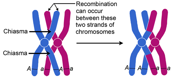
During prophase I, chromosomal condensation allows chromosomes to be viewed under the microscope. In late prophase I, homologous chromosomes (also called bivalent chromosomes, or bivalents) pair laterally, or side-by-side. At this time they are said to be in synapsis. During synapsis, crossovers – cross-connections that form from breakage and rejoining between sister chromatids – can occur between the paired bivalents, leading to genetic recombination (exchange of genetic material) between the strands involved. The point where a crossover occurs is called a chiasma (plural chiasmata) (see below figure). In figure below, following crossing over, the blue and red chromosomes, which originally carried AA and aa alleles, respectively, now carry Aa alleles in both chromosomes at the end of prophase I. Note that these bivalents have two chromosomes and four chromatids, with one chromosome originating from each parent.
Metaphase I
In metaphase I, each pair of bivalents (two chromosomes, four chromatids total) align on the metaphase plate. This is different from metaphase in mitosis, where all chromosomes align single file on the metaphase plate. The position of each chromosome in the bivalents is random - either parental homolog can appear on each side. This means that there is a 50-50 chance for the daughter cells to get either the mother's or father's homolog for each chromosome (see figure below). As shown in the below figure, during metaphase I, bivalents from either parent can align on either side of the cell. In an organism with two sets of chromosomes, there are four ways in which the chromosomes can be arranged, resulting in differences in chromosomal distribution in daughter cells after meiosis I. (A diploid organism with 2n chromosomes will have 2n possible combinations or ways of arranging its chromosomes during metaphase I.)
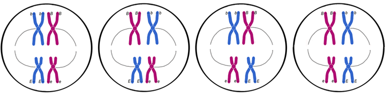
In a diploid cell with 2 pairs of chromosomes, there are 4 ways to arrange the chromosomes during metaphase I.
Anaphase I
In anaphase I, homologous chromosomes separate. Homologous chromosomes, each containing two chromatids, move to separate poles. Unlike in mitosis, the centromeres do not split and sister chromatids remain paired in anaphase I.
Telophase I and Cytokinesis
In telophase I, the homologs of each bivalent arrive at opposite poles of the cell, and a new nuclear membrane forms around each set of chromosomes. Cytokinesis then divides the cell into two daughter cells. Each of the two daughter cells is now haploid (n), with half the number of chromosomes per nucleus as in meiosis I. In some species, the nuclear membrane briefly forms around the chromosomes, while in others it does not. The cell now proceeds into meiosis II, with the chromosomes remaining condensed.